Trephine Surgery Steps in Pictures
Minimally invasive surgery is designed around the unique anatomy of the pilonidal unit: a skin opening (1) that leads to a narrow canal (2), which in turn extends into a subcutaneous pocket (3) containing inflammatory material and hair. Pilonidal disease may involve a single unit or several, and in some cases their pockets merge into one shared cavity..
During the procedure, each skin opening and its canal are excised with a trephine. This creates a wider channel with no skin lining, which allows the cavity to be cleaned effectively and to continue draining until it shrinks and closes, together with the skin opening.

Several pilonidal skin openings lead to a shared subcutaneous cavity, with a fistula and an additional skin opening on the right.

Structure of a pilonidal unit:
-
An opening in the skin of the natal cleft.
-
A tract partially lined with skin.
-
A pilonidal pocket containing hair.
How is the treatment performed?

Excision of the narrow pilonidal tract, partially lined with skin, using a trephine.

The widened tract allows access to the pilonidal pocket.

Cleaning of the pilonidal cyst using a surgical curette
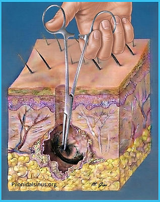
Cleaning of the pilonidal cyst using surgical forceps

The tract and the pilonidal pocket gradually shrink and disappear after cleaning